As presented by Dr. Osterhouse, Radiology in Practice teaches how to take and interpret radiographs, MRIs, CTs, and other diagnostic imaging tools, including the bone scan. In addition, the course provides a valuable review of spinal and extremity imaging for a doctor in general practice.
In this hour, Dr. Osterhouse reviews the x-ray physics to help the practitioner understand how to take better diagnostic films. She teaches how to mark films to aid in the diagnosis and management of spinal trauma and disease. She reviews the subject of rheumatoid arthritis and offers criteria for diagnosis. She also explains how and when to utilize flexion and extension stress film radiology in practice.
Learning Objectives
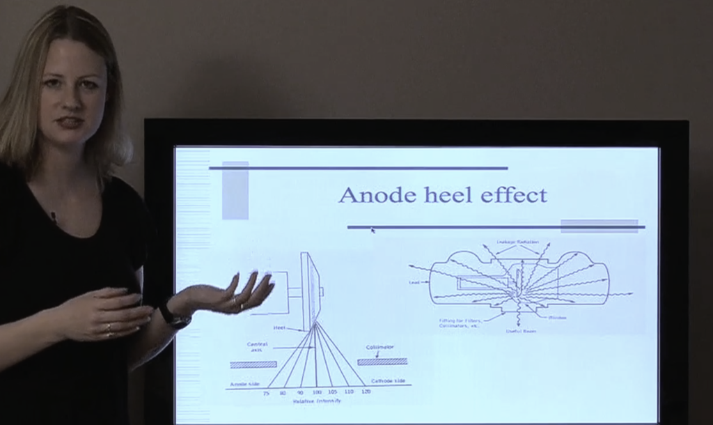
The participant will be able to explain how the x-ray beam is produced, how filters lower the patient’s radiation exposure, and how to consider the principles of x-ray physics to improve the quality of diagnostic images taken in the office. In addition, participants will be able to diagnose rheumatoid arthritis from plane films, mark films to measure the spinal curvatures, and explain when it would be appropriate to take flexion-extension plane film studies.